Dr. Morgan
Callahan
Board-Certified Veterinary Dentist
Endoscopic subgingival procedures. Surgical extractions under microscope. Feline stomatitis management. Complex oral cases that general practice can't crack — resolved, documented, and sent home the same day.
Accepting GP referrals · Second opinions welcome · Same-week appointments
Cases we see.
Explained plainly.
Each section pairs the actual radiograph or intraoral photograph with a plain-language clinical explanation — the same conversation we have with referring veterinarians every day.
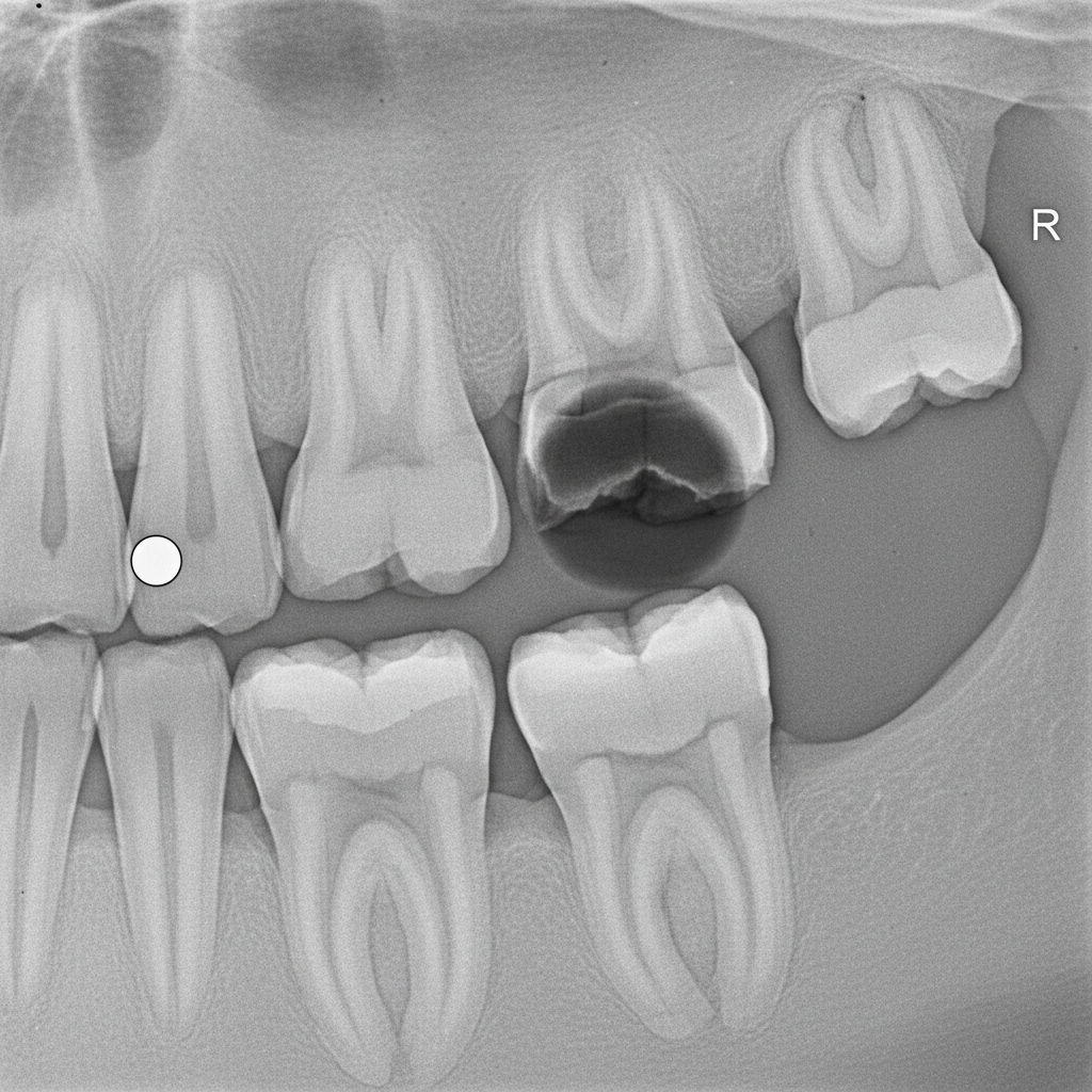
Periapical lucency confirms pulp necrosis — extraction indicated
The Tooth You Almost Missed
See this dark line running from the crown toward the root apex? That's a slab fracture on the upper fourth premolar — the carnassial. It looks like a minor chip on exam, but the pulp is exposed and the tooth has been dying for months.
General practice extractions often leave the palatal root behind. We use a surgical microscope and intraoral radiographs at every stage to confirm complete removal and rule out periapical pathology before closure.
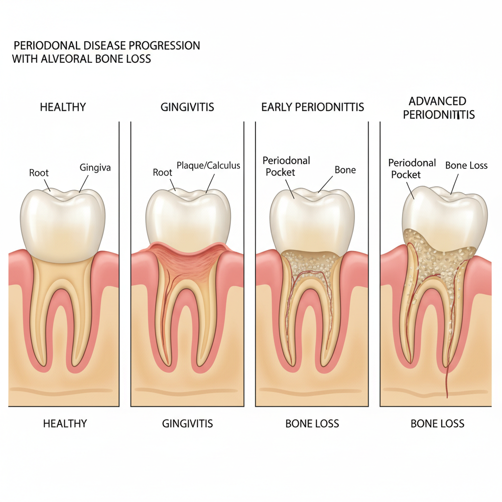
Bone loss >50% root length — surgical periodontal therapy vs. extraction
Stage III Isn't a Cleaning Problem
At Stage III, bone loss has already consumed more than 50% of the root support. A polish and scale won't reverse this. The question is which teeth are salvageable and which are causing occult pain your patient can't tell you about. We probe every pocket, radiograph every root, and stage each tooth individually. Owners often say their pet "started eating again" post-op — because they didn't know it hurt until it stopped.
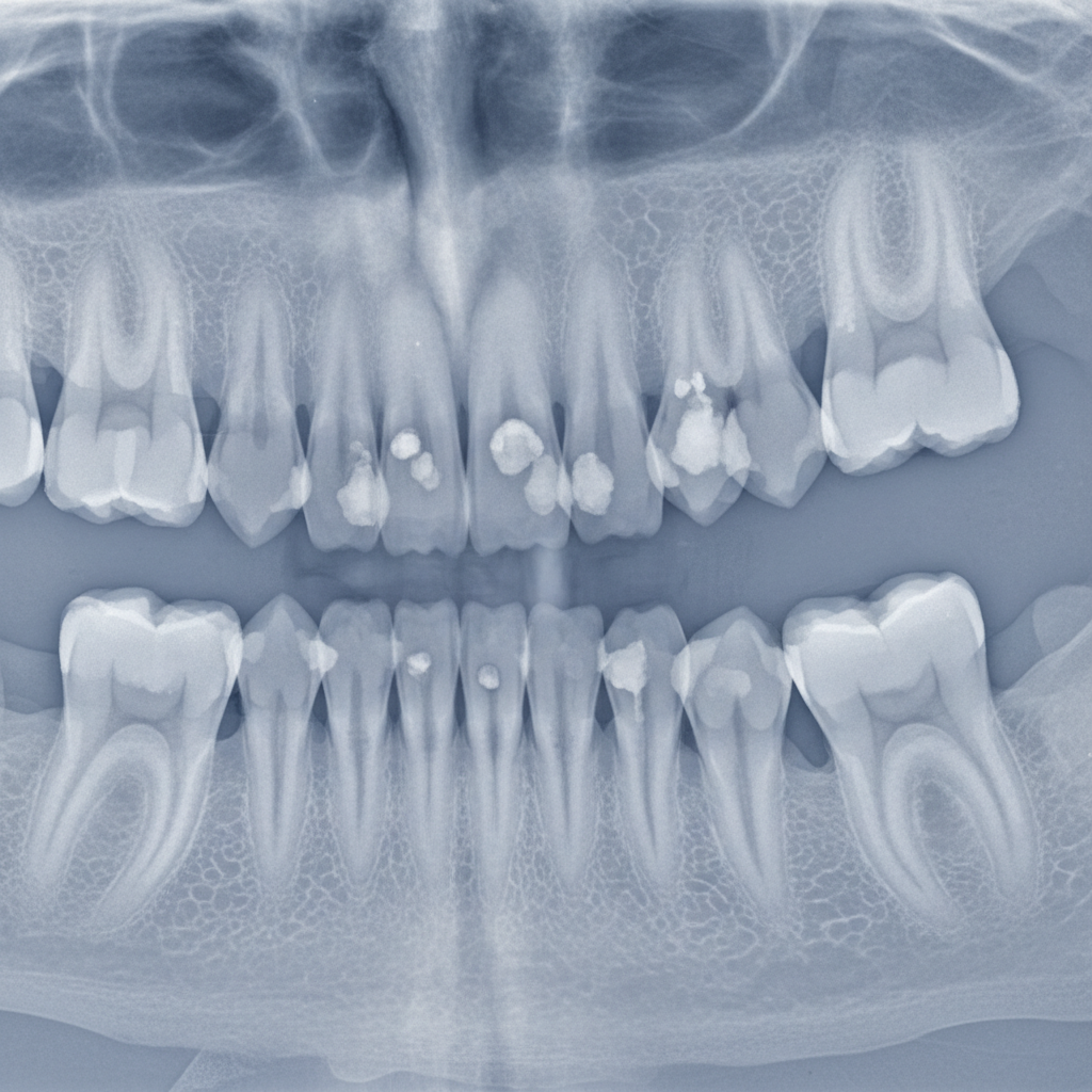
Root replacement resorption — crown amputation protocol appropriate
The Cavity That Isn't a Cavity
Type 2 feline resorptive lesions show root replacement resorption — the body is actively converting tooth structure into bone-like tissue. These are not caries. Attempting standard extraction shears the root and leaves fragments.
We identify lesion type radiographically before every extraction. Type 2 lesions are managed with intentional root retention and crown amputation — a technique that eliminates pain while avoiding iatrogenic fracture.
Three referral paths.
One specialist.
Whether you're a general practitioner with a complex case, a pet owner seeking a second opinion, or a shelter managing chronic oral disease — there's a direct path here.
You diagnosed it. We'll resolve it.
Complex extractions, intraoral radiograph interpretation, pre-surgical staging, and post-op notes returned within 24 hours. We keep you in the loop and send them back to you for routine care.
Referral packets · Surgical reports · Continuing education
Download Referral GuideA second opinion before extraction.
If your veterinarian has recommended extracting a tooth and you want another perspective, we offer consultation appointments with intraoral radiographs and a written treatment plan. Extraction is sometimes right. But not always.
Consultations · Written reports · Treatment alternatives
Check Symptoms FirstStomatitis cats deserve a path forward.
Full-mouth extraction with documented post-op protocols, adoption-ready dental reports, and volume scheduling for multi-cat intakes. We've helped cats that cycled through four foster homes find permanent placement after resolution.
Volume scheduling · Adoption reports · Foster protocols
Speak With Our TeamThe Fang Referral
Clinical Guide
A 24-page clinical reference built for general practitioners — covering referral criteria, radiograph interpretation, and post-op protocols. Updated February 2026.
- Referral criteria by case type
- Intraoral radiograph interpretation guide
- Feline stomatitis staging protocol
- Pre-surgical patient prep checklist
- Post-op care instructions (printable)
- Contact & emergency consultation line
PDF · 24 pages · 3.2 MB
Printable · AVDC-reviewed content
Download the guide
Enter your practice name and email — we'll send the PDF immediately and follow up only if you want consultation support.
No marketing emails. One delivery confirmation, one follow-up offer — that's it. Unsubscribe instantly.
Check your pet's oral symptoms
Select every symptom you've observed. This isn't a diagnosis — it's a structured starting point for a conversation with a specialist.
Receive a personalized
oral health summary
We'll email you a plain-language summary of the symptoms you selected, what they may indicate, and whether a specialist consultation is recommended.
This tool does not provide a diagnosis. Results are educational and for discussion purposes only.
Ready to book?
(415) 882-3940
Mon–Fri · 8am–5pm PT